
X-Ray
Most dental problems don’t exist only on the surface. Pain, infection, bone changes, and alignment issues often develop beneath the gums long before they become obvious during a visual exam. Dental imaging allows us to see what cannot be seen directly so decisions are made based on evidence, not assumptions. At KSV Dental, X-rays are used to confirm a diagnosis, guide treatment, and prevent surprises, not as a routine step applied to every patient. The goal is clarity before action.

Why Imaging Is Taken Before Treatment
Without imaging, treatment planning becomes guesswork. Small issues can be underestimated, and complex ones can be missed entirely.
Dental X-rays help:
Identify hidden decay or infection
Assess bone levels and tooth roots
Plan treatments accurately
Avoid unnecessary or incomplete procedures
Imaging is selected based on what information is needed — no more, no less.
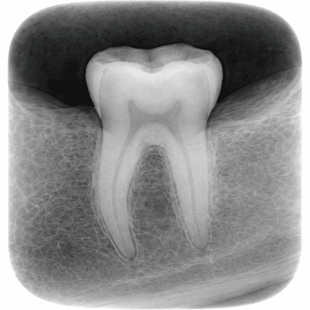
Periapical (P.A) X-Ray: Focused Detail Where It Matters
Periapical X-rays provide a close-up view of a single tooth, including its root and surrounding bone. This type of imaging is commonly used when a specific tooth is causing symptoms.
P.A X-rays are useful for:
Diagnosing tooth infections
Evaluating root canal conditions
Monitoring healing after treatment
They offer precise detail in a targeted area, making them efficient and informative.
OPG (Panoramic X-Ray): The Full Overview
An OPG provides a wide view of the entire mouth, including all teeth, jawbones, and surrounding structures.
This scan is often used to:
Assess overall oral health
Identify impacted teeth
Evaluate jawbone condition
Support treatment planning across multiple areas
The panoramic view helps place individual issues into context before deciding on next steps.
Cephalometric X-Rays: Understanding Structure and Alignment
Cephalometric imaging captures the relationship between teeth, jaws, and facial structure.
Lateral Cephalometric
Used primarily in orthodontic planning, this view helps assess bite alignment and jaw position from the side.
Frontal Cephalometric
This view provides symmetry and alignment information from the front, supporting balanced treatment planning.
These images are not taken routinely — they are used when structural analysis is needed to guide orthodontic or corrective care.
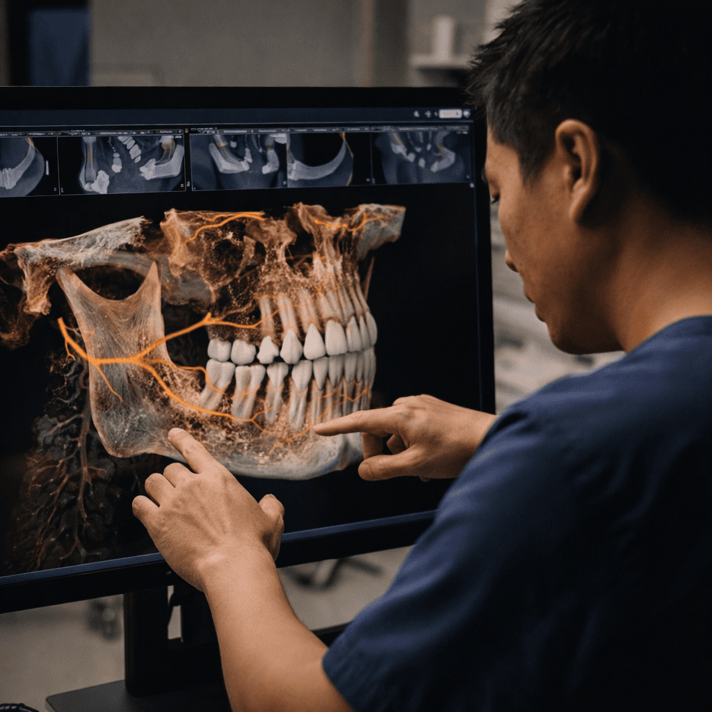
3D CBCT: Advanced Imaging for Complex Decisions
Cone Beam Computed Tomography (CBCT) creates a three-dimensional view of teeth, bone, nerves, and surrounding structures.
At KSV Dental, CBCT imaging is available in-house and used selectively when conventional X-rays do not provide enough information.
CBCT may be recommended for:
Implant planning
Complex extractions
Root canal complications
Detailed assessment near nerves or sinuses
This level of detail improves accuracy and reduces risk — especially in complex cases.
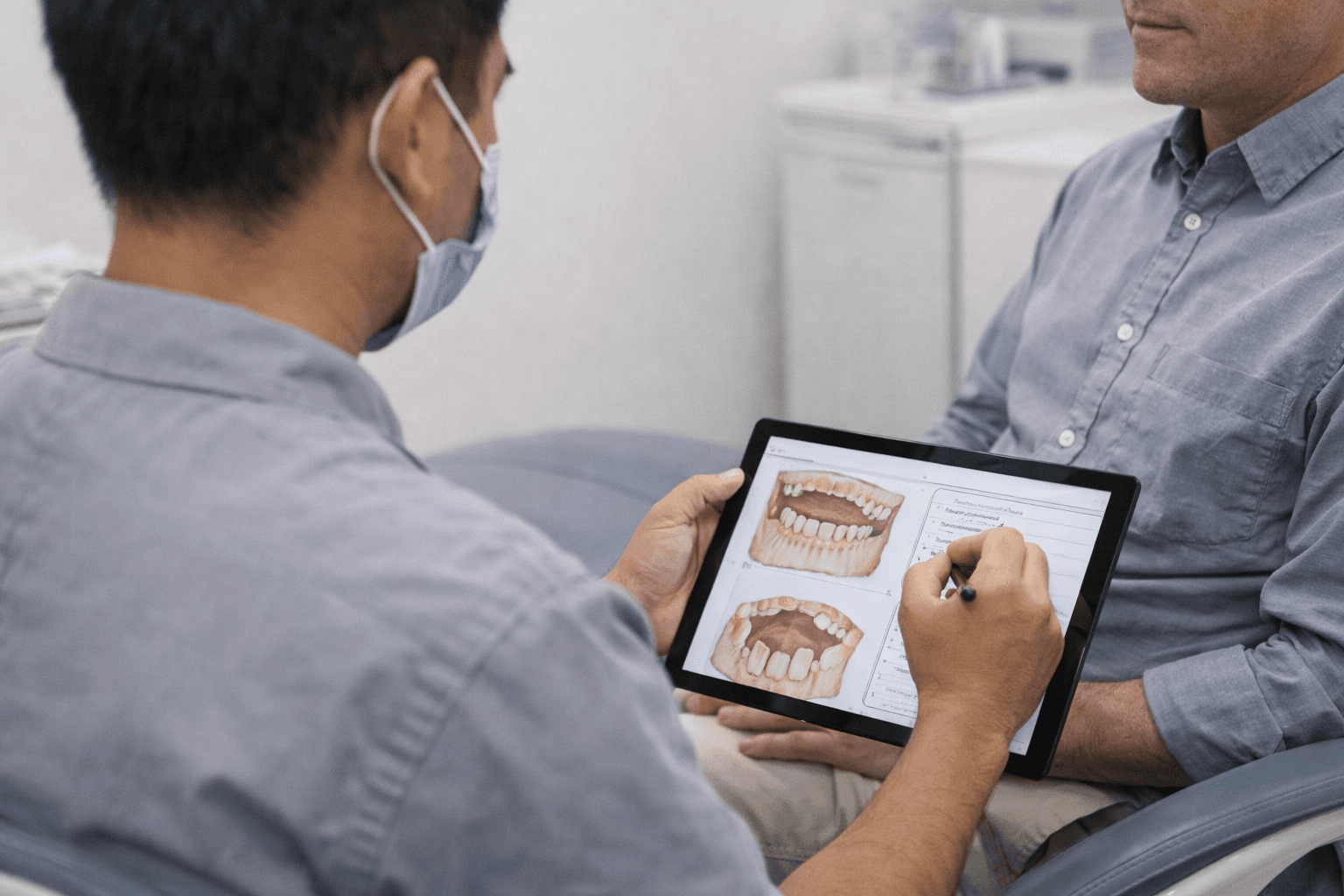
Imaging Chosen With Purpose, Not Habit
Not every patient needs every type of scan.
At KSV Dental, imaging is chosen based on:
Symptoms and clinical findings
Treatment being considered
The information required to make a safe decision
If a scan isn’t necessary, it isn’t taken. If it adds clarity or reduces uncertainty, it’s explained clearly before proceeding.
This approach avoids unnecessary exposure and keeps care proportional.
Safety and Radiation Considerations
Modern dental imaging uses low-dose radiation, and every scan is performed with safety in mind.
The benefit of accurate diagnosis and proper planning far outweighs the minimal exposure involved. Imaging is taken thoughtfully, not repeatedly or casually.
Better Imaging Means Fewer Surprises
Accurate imaging helps:
Reduce unexpected findings during treatment
Shorten overall treatment time
Improve predictability of outcomes
When the full picture is clear from the start, treatment proceeds more smoothly.
A Foundation for Confident Decisions
Dental imaging is not about doing more — it’s about doing things right.
At KSV Dental, X-rays and scans support careful diagnosis, precise planning, and informed decisions. They ensure treatment is guided by evidence, not guesswork.
Seeing clearly first is what allows everything else to be done properly.
Why Imaging Is Taken Before Treatment
Without imaging, treatment planning becomes guesswork. Small issues can be underestimated, and complex ones can be missed entirely.
Dental X-rays help:
Identify hidden decay or infection
Assess bone levels and tooth roots
Plan treatments accurately
Avoid unnecessary or incomplete procedures
Imaging is selected based on what information is needed — no more, no less.
Periapical (P.A) X-Ray: Focused Detail Where It Matters
Periapical X-rays provide a close-up view of a single tooth, including its root and surrounding bone. This type of imaging is commonly used when a specific tooth is causing symptoms.
P.A X-rays are useful for:
Diagnosing tooth infections
Evaluating root canal conditions
Monitoring healing after treatment
They offer precise detail in a targeted area, making them efficient and informative.
OPG (Panoramic X-Ray): The Full Overview
An OPG provides a wide view of the entire mouth, including all teeth, jawbones, and surrounding structures.
This scan is often used to:
Assess overall oral health
Identify impacted teeth
Evaluate jawbone condition
Support treatment planning across multiple areas
The panoramic view helps place individual issues into context before deciding on next steps.
Cephalometric X-Rays: Understanding Structure and Alignment
Cephalometric imaging captures the relationship between teeth, jaws, and facial structure.
Lateral Cephalometric
Used primarily in orthodontic planning, this view helps assess bite alignment and jaw position from the side.
Frontal Cephalometric
This view provides symmetry and alignment information from the front, supporting balanced treatment planning.
These images are not taken routinely — they are used when structural analysis is needed to guide orthodontic or corrective care.
3D CBCT: Advanced Imaging for Complex Decisions
Cone Beam Computed Tomography (CBCT) creates a three-dimensional view of teeth, bone, nerves, and surrounding structures.
At KSV Dental, CBCT imaging is available in-house and used selectively when conventional X-rays do not provide enough information.
CBCT may be recommended for:
Implant planning
Complex extractions
Root canal complications
Detailed assessment near nerves or sinuses
This level of detail improves accuracy and reduces risk — especially in complex cases.
Imaging Chosen With Purpose, Not Habit
Not every patient needs every type of scan.
At KSV Dental, imaging is chosen based on:
Symptoms and clinical findings
Treatment being considered
The information required to make a safe decision
If a scan isn’t necessary, it isn’t taken. If it adds clarity or reduces uncertainty, it’s explained clearly before proceeding.
This approach avoids unnecessary exposure and keeps care proportional.
Safety and Radiation Considerations
Modern dental imaging uses low-dose radiation, and every scan is performed with safety in mind.
The benefit of accurate diagnosis and proper planning far outweighs the minimal exposure involved. Imaging is taken thoughtfully, not repeatedly or casually.
Better Imaging Means Fewer Surprises
Accurate imaging helps:
Reduce unexpected findings during treatment
Shorten overall treatment time
Improve predictability of outcomes
When the full picture is clear from the start, treatment proceeds more smoothly.
A Foundation for Confident Decisions
Dental imaging is not about doing more — it’s about doing things right.
At KSV Dental, X-rays and scans support careful diagnosis, precise planning, and informed decisions. They ensure treatment is guided by evidence, not guesswork.
Seeing clearly first is what allows everything else to be done properly.
More Service
More Service
Other specific dental treatments we offer
Other specific dental treatments we offer


Opening Hours
Monday - Friday
08:30AM - 12:00PM, 01:00PM - 6:00PM
Saturday
08:00AM - 12:00PM, 01:00PM - 6:00PM
Sunday
08:00AM - 02:00PM
Service Location
Opening Hours
Monday - Friday
08:30AM - 12:00PM, 01:00PM - 6:00PM
Saturday
08:00AM - 12:00PM, 01:00PM - 6:00PM
Sunday
08:00AM - 02:00PM
Service Location
Opening Hours
Monday - Friday
08:30AM - 12:00PM, 01:00PM - 6:00PM
Saturday
08:00AM - 12:00PM, 01:00PM - 6:00PM
Sunday
08:00AM - 02:00PM
Service Location